Campylobacter hund
Send a Message
This message will be pushed to the admin's iPhone instantly.
 Campylobacter bacteria are important causes of disease in people. Many Campylobacter species exist, and these different species vary quite a bit in their ability to cause disease in people and animals. Campylobacter jejuni is one of the most common causes of diarrhea in people worldwide, and is most commonly associated with contaminated food. However, a few studies have reported that having pets (especially pets with diarrhea) is also a risk factor for Campylobacter jejuni infection.
Another Campylobacter species that may be of concern is Campylobacter upsaliensis. This species is primarily associated with dogs and cats, and a large percentage of healthy dogs and cats may be shedding this bacterium in their stool at any time. It doesn’t seem to be a cause of disease in dogs and cats, but it may be an important and overlooked cause of disease in people. One study from the US reported that C. upsaliensis was the 2nd most common Campylobacter strain found in people with diarrhea (after C. jejuni). However, the true role of this species is unclear, partly because of common laboratory testing methods. Culture is the main method used to diagnose infection with Campylobacter, but this bacterium can be difficult to grow in the lab. Usually, culture media for Campylobacter contain antibiotics to inhibit other better/faster growing bacteria. Unfortunately, C. upsaliensis is often inhibited by these antibiotics, so it’s likely to be missed in these cases even if it is there. Therefore, we might be underestimating the role of this Campylobacter species in diarrhea. This is an critical issue to investigate because C. upsaliensis is so common in dogs and cats, and it’s important to determine what role pets play in human disease.
Avoiding Campylobacter infection involves some basic steps: avoid contact with feces, take care when handling diarrhea from pets, wash your hands regularly after handling pets and always wash your hands thoroughly after any contact with feces. Make sure your physician knows you have pets. In particular, if you have a pet with diarrhea or have recently acquired a new pet (especially a puppy or kitten), make sure Campylobacter infection is considered if you get diarrhea. Most infections are mild and go away on their own but some require specific treatment.
More information about Campylobacter can be found on the Worms & Germs Resources page.
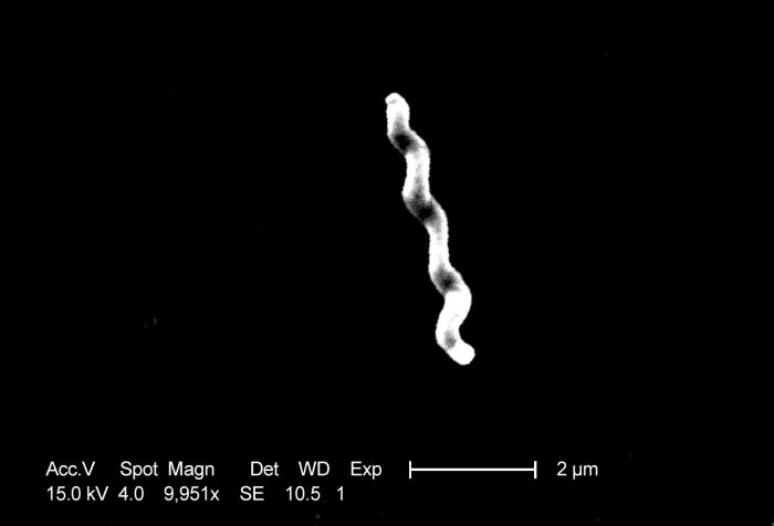
Image credit: CDC/ Dr. Patricia Fields, Dr. Collette Fitzgerald
Copyright University of Guelph. All Rights Reserved.
Magsjuk av kyckling (Campylobacter)

Bakterierna campylobakter finns i otillagad kyckling och kan orsaka kräkningar och diarré.
Campylobacter är en bakterie som orsakar mag- och tarmsymtom. Den är nära besläktad med salmonellabakterien, och orsakar magsmärtor, illamående, kräkningar, diarré och feber. Den vanligaste smittokällan är vid konsumtion av dåligt tillagad kyckling.
Omkring 8 000 svenskar drabbas varje år, och då smittas häften inom Sverige och resten utomlands. Detta gör campylobakter till den bakterie som orsakar flest fall av magsjuka i landet. Statistik visar en tydlig ökning under semestermånaderna juni, juli och augusti. Det finns i nuläget inget vaccin mot sjukdomen, och några av de bästa sätten att skydda sig från att bli magsjuk av kyckling är genom att undvika otillagad mat och att vara noga med hygien och sanitet när man handskas med rå kyckling. Campylobacter anses som en allmänfarlig sjukdom, och alla sjukdomsfall ska rapporteras till Folkhälsomyndigheten.
Varför kan man bli magsjuk av kyckling?
Kyckling är ett mycket populärt livsmedel både i Sverige och utomlands, men det är viktigt att se till att man inte får i sig rått kycklingkött. Livsmedelsverket har genomfört en undersökning som visar att var femte slaktad kyckling innehåller smittan. Det krävs mycket lite av bakterien för att en människa ska bli magsjuk av kyckling, vilket gör rått kycklingkött till en stor hälsofara. Om kycklingköttet är ordentligt tillagat så finns dock ingen risk att man blir sjuk. Däremot är det viktigt att tänka på att diska bestick och skärbrädor som har kommit i kontakt med rått kött, då även detta kan vara en smittokälla. Bakterierna kan överleva i en bärare (så som kyckling) i upp till tre veckor, och även om längre överlevnadstid är ovanligt i Sverige så förekommer det. Även andra sorters kött kan innehålla smittan, om än inte i samma utsträckning. Levande hundar, katter och vilda djur kan också vara bärare, men insjuknar sällan själva.
Symtom när man blir magsjuk av kyckling
De flesta insjuknar 1-3 dagar efter smittotillfället, men det kan i vissa fall ta upp till 10 dagar. Ofta insjuknar man oväntat och utan förvarning. Symtom på att man blivit magsjuk av kyckling kan vara diarré (som ibland är blodblandad), intensiva magsmärtor, illamående, kräkningar och feber. Ledbesvär förekommer också i viss utsträckning. I svårare fall av smittan så kan man uppleva frossa och utmattning.
Diagnosen ställs med hjälp av ett avföringsprov. Det görs en bakterieodling på provet, och en läkare kan sedan avgöra om det är Campylobacter som orsakar magsjukan.
Behandling
De flesta fall av smittan kräver ingen läkarvård, och man blir oftast frisk igen inom två veckor. Smittan kan dock leva kvar i avföringen i upp till fem veckor. Det är viktigt att dricka mycket vatten för att ersätta det som kroppen har förlorat, och att vila. Vissa läkare rekommenderar att man blandar i lite socker i vattnet, samt att man dricker det i små mängder åt gången. Om infektionen däremot sprider sig till blodet, vilket är när man kan uppleva frossa, så bör den behandlas. En kur av antibiotika skrivs då ut för att påskynda tillfrisknandet
Övrigt om magsjuka orsakat av kyckling och campylobakter
När man är utomlands så kan det vara bra att vara extra försiktig, då smittan förekommer mer i vissa andra länder än vad den gör i Sverige. Om man går ut på restaurang och är osäker på hur kycklingen tillagas så kan det vara lika bra att undvika kyckling helt och hållet.
Volksleiden Darm: Darmbeschwerden und Darmprobleme
Informationen aus der Naturheilpraxis von René Gräber

Der bekannte Fastenarzt Franz Xaver Mayr ( F.X. Mayr Kur) sagt zu den Darmproblemen der Neuzeit: Chronische Darmstörungen sind das unbekannteste, weitverbreitetste und verhängnisvollste Leiden des modernen Menschen".
Und in der Tat erzählen Witze von den Lieblingsthemen älterer Menschen: Essen, Wetter, Krankheiten und der letztmalige Stuhlgang.Der Magen-Darm-Trakt ist ein komplexes Organ und kann unter Umständen sehr störanfällig sein. Daneben gibt es einige Erkrankungen, die das Verdauungssystem betreffen.
Häufige Erkrankungen des Darms
Häufig ist der Reizdarm bzw. der Reizmagen. Bei diesen Krankheitsbildern kommt es in bestimmten Situationen oder immer wiederkehrend zu Verdauungsunregelmäßigkeiten mit Bauchschmerzen und Blähungen. Eine "organische Ursache" für die Beschwerden kann beim Reizdarm- bzw. Reizmagen-Syndrom durch die meisten Schulmediziner nicht gefunden werden;
Diese Erkrankungen gehören für die meisten Ärzte zu den "funktionellen Störungen", bei denen eine psychosomatische Ursache vermutet wird.
Vornehmlich im Kindesalter, aber auch in allen anderen Altersklassen, kann es zu einer Infektion des Magen-Darm-Trakts mit Bakterien, Viren, Pilzen oder Parasiten kommen (Magen-Darm-Grippe = Gastroenteritis).
Häufig findet man hier Noro-Viren oder Rotaviren, das Bakterium Campylobacter jejuni, welches vor allem durch nicht durchgebratenes Geflügelfleisch übertragen wird, Salmonellen (Salmonelleninfektion vor allem über Eier und Eiprodukte), den Pilz Candida albicans oder Würmer und Amöben.
Die Symptome sind Гњbelkeit, Durchfall und Bauchschmerzen; auch Reisedurchfall wird durch Infektionen verursacht.
Neben den Nahrungsmittelunverträglichkeiten und Nahrungsmittelallergien (z.B. Laktoseintoleranz, Zöliakie) können auch chronisch entzündliche Darmerkrankungen (z.B. Colitis ulcerosa, Morbus Crohn) Durchfälle und Bauchschmerzen verursachen.
Ebenfalls häufig ist die Refluxkrankheit (gastroösophageale Refluxkrankheit). Hierbei kommt es durch eine Funktionsstörung des unteren Speiseröhrenschließmuskels zu einem Rückfluss von saurem Magensaft in die Speiseröhre (Ösophagus). Das führt zu Sodbrennen vor allem nach großen Mahlzeiten, im Liegen oder beim Vornüberbeugen. Außerdem kann es zu Oberbauchschmerzen kommen. Die chronische unbehandelte Refluxkrankheit kann zu Husten, Kehlkopfentzündung oder Asthma führen und erhöht das Risiko, an einem Speiseröhrenkrebs (Ösophaguscarcinom) zu erkranken.
Auch häufig kommt es zu Magenschleimhautreizungen und Magenschleimhautentzündungen sowie einem Magengeschwür und Zwölffingerdarmgeschwüren (Ulcus ventriculi bzw. Ulcus duodeni). Eine Magenschleimhautentzündung (Gastritis) kann durch eine dauerhafte Einnahme von Schmerzmedikamenten (z.B. ASS, Ibuprofen), Rauchen, übermäßigen Alkoholkonsum oder Stress entstehen.
Aber auch Autoimmunerkrankungen oder die Infektion der Magenschleimhaut mit dem Bakterium Helicobacter pylori können eine Gastritis hervorrufen. Bei einer chronischen Gastritis steigt das Risiko ein Magen- oder Zwölffingerdarmgeschwür zu bekommen sowie das Risiko einen Magenkrebs (Magencarcinom) zu entwickeln.
Blinddarmreizungen und Blinddarmentzündungen (genauer eine Entzündung des Wurmfortsatzes = Appendizitis) treten vor allem im Kindes- und jungen Erwachsenenalter auf und äußern sich durch Übelkeit, Erbrechen, eine erhöhte Temperatur und Bauchschmerzen (im Krankheitsverlauf im rechten Unterbauch lokalisiert). Wird die Blinddarmentzündung nicht rechtzeitig diagnostiziert und behandelt, kann es zu ernsthaften Komplikationen mit Todesfolge kommen.
Die Divertikulitis wird oft als Appendizitis des älteren Menschen bezeichnet. Dabei entzünden sich Darmausstülpungen (Darmdivertikel) des Dickdarms und verursachen Schmerzen, die im linken Unterbauch lokalisiert sind. Hinzu können Stuhlunregelmäßigkeiten kommen.
Bei fast jedem Menschen findet man Hämorrhoiden. Das sind Gefäßaussackungen im Analbereich, die die Kontinenz auch für Darmgase gewährleisten. Hämorrhoiden werden zu einer Erkrankung, wenn der Blutabfluss aus dem Gefäßpolster nicht mehr möglich ist und eine dauerhafte Schwellung besteht. Die Schleimhaut kann an dieser Stelle einreißen, es kommt zu Blutauflagerungen auf dem Stuhl und am Toilettenpapier, im Krankheitsverlauf kann es zu Jucken, Brennen und Nässen kommen.
Darmkrebs betrifft vor allem ältere Menschen und kommt häufiger im Dick- als im Dünndarm vor. Erste Anzeichen können Stuhlunregelmäßigkeiten und verdeckte (okkulte) Blutbeimengungen sein. Später ist sogar ein Darmverschluss (Ileus) möglich. Eine Vorstufe von Darmkrebs können Darmpolypen (Schleimhautwucherungen) sein.
Sprache der Symptome
Es gibt eine ganz einfache Frage zu den Darmbeschwerden: Gibt es etwas, was Sie einfach nicht verdauen können?
Weitere mögliche Ursachen
Die Haupt-Ursache für jegliche Art von Darmproblemen und Darmbeschwerden sind grundsätzlich immer falsche Ernährung, Bewegungsmangel und Stress.

Abb.1: Das Sprichwort: "Du bist was Du iГџt", gilt auch fГјr die allermeisten Darmbeschwerden.
NatГјrlich sind die LehrbГјcher zu den verschiedensten Darmproblemen lang und fГјr eine spezifische naturheilkundliche oder alternative Therapie ist eine genauere Diagnose wichtig.
Ein weitere Störquelle ist das latente Problem der Darmflora, die durch Medikamente (vor allem Antibiotika) und falsche Ernährung stark beeinflusst wird.
Гњbrigens: Wenn Sie solche Informationen interessieren, dann fordern Sie doch einfach meinen kostenlosen Praxis-Newsletter an:
Im Folgenden finden Sie naturheilkundliche und Alternative Therapieverfahren, die bei Darmbeschwerden in Frage kommen können.
Es gibt viele Dinge, die ich empfehle zu meiden. Vor allem aber Schweinefleisch, Milch und Eier.
Bei den Heilverfahren liegt das Fasten eindeutig vorne - bei fast allen Darmbeschwerden. Allgemein sei hier nur auf eine gesunde Ernährung verwiesen. Patienten rate ich generell auch erst einmal zu einer Schonkost. Wie diese aussehen kann, beschreibe ich im Beitrag: Schonkost - Eine Anleitung für Patienten.
Resistente Stärke kann die Darmgesundheit fördern.
Die FuГџreflexzonenmassage eignet sich, wenn die Darmprobleme vor allem durch Stress verschlechtert werden. Das erkennt man daran, dass die Symptome alle besser sind, wenn man Urlaub hat. Ich hatte Patienten, die ihre funktionellen Darmprobleme (ohne weitere Befunde) lediglich mit dieser Therapie Гјber die FuГџreflexzonen gut im Griff hatten.
Es gibt unzählige Heilpflanzen die helfen. Da kommt es aber auf die spezifischen Beschwerden an.
Lactobacillus acidophilus verbessert nachweislich die Verdauungsfunktion. DarГјberhinaus wirkt L. acidophilus bei Durchfall in Folge Antibiotika.
Bei den SchГјssler Salzen kommen in Frage:
- akuter Magen-Darm-Katarrh: Ferrum phosphoricum
- Magen-Darm-SchleimhautentzГјndung mit Magenschmerzen und Erbrechen von Schleim: Natrium chloratum
- Magen-Darm-Schleimhautentzündung mit trockener Zunge und körperlicher Schwäche: Kalium phosphoricum
- Magen-Darm-SchleimhautentzГјndung, mit krampfartigen Magenschmerzen ohne Fieber, reine Zunge: Magnesium phosphoricum
Grundsätzlich empfehle ich bei jeglicher Art von Darmbeschwerden mehr Bewegung und verordne wenigstens einen täglichen Spaziergang.
Zur Stimulierung der Verdauung ist nämlich die Bewegung der Eingeweide wichtig und dazu trägt die Erschütterung des Gehens und Laufens bei.
Der Effekt lässt sich mit einem sog. Rebound (Minitrampolin) vervielfachen. Eine Minute leichtes Hüpfen entspricht fünf Minuten gehen. Hüpfen oder wippen Sie täglich zweimal drei bis fünf Minuten.
Und zum Schluß vielleicht noch etwas, von dem Sie wahrscheinlich noch nie gehört haben und das auch etwas "kurios" anmutet: Das Ano-Darmpessar. Richtig und regelmäßig angewendet, kann dieses Röhrchen dazu beitragen Darmprobleme dauerhaft zu reduzieren.
Dieser Beitrag wurde letztmalig am 2.4.2016 aktualisiert.
Campylobacteriose
Af dyrlæge Peter Kierk
Hunde og katte kan blive inficeret med denne tarmbakterie, men mange andre pattedyr, herunder også mennesker, kan også få infektionen. En infektion, som kan smitte fra dyr til mennesker kaldes en zoonose. Campylobacteriose findes overalt på jordkloden og spredes via afføring fra inficerede dyr.
Behandlingen af dyret fortsætter, til der ved en fornyet dyrkning ikke kan påvises campylobacter i afføringen. Da man flere gange har påvist campylobacter i mælk og i kød fra specielt fjerkræ, er man i dag meget opmærksom på bakterien. Mennesker kan nemlig blive alvorligt syge ved indtagelse af inficerede fødevarer og ved omgang med smittede hunde og katte. Det kan derfor ikke understreges nok, at hygiejne, tilstrækkelig varmebehandling af kød herunder fjerkræ, etc. er vigtige foranstaltninger for at undgå smittespredning.
Prognosen for et dyr, der bliver korrekt behandlet for campylobacteriose, er god.
Forskningen af campylobacters betydning hos hunde og katte er først rigtig kommet i gang inden for de sidste ti år, og der er stadig en del ubesvarede spørgsmål omkring denne zoonose.
Diarré er et symptom der beskriver øget frekvens, mængde og/eller en mere vandig konsistens af afføringen.Diarré opstår når tarmens evne til at optage væske fra tarmens indre hindres. Diarré kan også …
Opkast er en kraftfuld udtømning af mavesækken, og evt. indholdet af den forreste del af tyndtarmen. Det er vigtigt at skelne mellem opkast regurgitation (passivt tilbageløb af indtaget foder og/…

Foder ved diarré og opkast, colitis eller dårlig fordøjelse

Genoprettelse af væske- og elektrolytbalancen hos hund og kat

Mavetarmdiæt til hunde

Tabletter til regulering af tarmfunktionen ved diarré

Fodertilskud til dårligt fungerende mave.

Pasta til genopretning af fordøjelsen

Kosttilskud til afhjælpelse af dårlig fordøjelse

Mavetarmdiæt til hund
Vi sender faglig information, tips og tricks og gode tilbud 2-4 gange om måneden. Nye abonnenter får en rabatkode på 10% til webshoppen.


 Indhold på Netdyredoktor.dk
Indholdet på Netdyredoktor.dk er udelukkende til informationsbrug. Disse informationer må på ingen måde kompensere eller erstatte den professionelle rådgivning og behandling, som gives af en autoriseret dyrlæge. Læs afsnittet om juridisk information og ansvarsfraskrivelse samt cookiepolitik .
Vores familiedyr er altid overladt til menneskers omsorg, både hvad angår pleje, sundhed og sygdom.
Det forpligter både dig som dyreejer og os som veterinærsygeplejersker og dyrlæger. Vi er med i et kompetent netværk, hvor vi altid kan trække på hinandens ekspertise. På den måde får dit dyr altid den bedste behandling.
Overview of Enteric Campylobacteriosis
By Alicja E. Lew-Tabor, BSc (Hons), PhD, Principal Research Fellow, Queensland Alliance for Agriculture & Food Innovation, The University of Queensland
- Enteric Campylobacteriosis
Campylobacter spp are spiral, microaerobic, gram-negative bacteria that cause gastroenteritis in people and animals. Several Campylobacter spp are zoonotic. Many domestic animals develop acute gastroenteritis after ingestion of Campylobacter spp, including dogs, cats, calves, sheep, pigs, ferrets, mink, monkeys, and several species of laboratory animals. (See also Bovine Genital Campylobacteriosis, see Zoonotic Diseases, and see Avian Campylobacter Infection.) Infection with C jejuni is one of the most common causes of gastroenteritis in people worldwide and is the most extensively studied Campylobacter species.
Campylobacter spp are spiral or curved rods that exhibit a characteristic corkscrew darting motility, mediated by a single polar flagellum. These are slow growing, with a generation time of
90 min, fastidious, and require enriched medium and microaerobic conditions with increased CO2 (3%–15% O2, 3%–10% CO2, 85% N2) for growth.
The family Campylobacteraceae consists of three genera, including Campylobacter and Arcobacter associated with animal and human diseases. Certain species are present commensally in animals as suspected reservoirs for human infections. The thermophilic Campylobacter spp, C jejuni, or C coli have the highest prevalence and disease impact. Campylobacter species causing diseases in livestock include C jejuni subsp jejuni (enteritis and abortion), C coli, C mucosalis (porcine enteritis), C upsaliensis, C helveticus (companion pet enteritis), C hyointestinalis subsp hyointestinalis (porcine and bovine enteritis), C sputorum (abortions in sheep), and C fetus subsp fetus (isolated from intestinal tracts of sheep and cattle, sporadic abortions). Certain species such as C jejuni, C hyointestinalis, and C fetus possess closely related subspecies with different disease foci. Initially, Arcobacter spp were considered to be aerotolerant campylobacters and are implicated in reproductive disorders, mastitis, gastric ulcers, and/or diarrhea in livestock, including A cryaerophilus (previously C cryaerophila), A skirrowii, A thereius, and A butzleri.
Transmission and Epidemiology:
Transmission is food- or waterborne or via fecal-oral spread. Animals serve as reservoir hosts for Campylobacter spp infections in both animals and people throughout the world. The predominant ecologic niche for Campylobacter spp is the GI tract of a wide variety of domesticated and wild vertebrates, and zoonotic transmission from animals to people in meat of animal origin, especially chicken, is a food safety issue. Campylobacter spp are also commonly isolated from free-living birds, including migratory birds and waterfowl, crows, gulls, and domestic pigeons, which can contaminate environments of grazing animals. Wild rodents and insects such as flies have also been reported to harbor and transmit C jejuni. Fecal contamination of the environment provides a ubiquitous source of these organisms under appropriate conditions for their survival. Campylobacter spp can persist for long periods in feces, milk, water, and urine, especially at temperatures close to 4ºC. In adverse conditions, C jejuni jejuni converts to a viable nonculturable form that can be reactivated when ingested.
Human foods documented as contaminated with Campylobacter include chicken, turkey, beef, pork, fish, and milk. Domesticated poultry are the most significant reservoir of C jejuni jejuni for people, causing 50%–70% of cases; chicken meat is the number one source. Dogs and cats are commonly infected similar to their owners when they ingest undercooked poultry.
Pathogenesis:
Bacterial motility, mucus colonization, toxin production, attachment, internalization, and translocation are among the processes associated with C jejuni jejuni virulence. Infection begins with ingestion of C jejuni jejuni in contaminated foods or water. Gastric acid provides a barrier, and the bacteria must reach the small and large intestines to multiply; C jejuni invades both epithelial cells and cells within the lamina propria.
Clinical Findings:
Abdominal pain, fever, diarrhea, blood in feces, and inflammatory cells in feces demonstrate the inflammatory nature of the infection. Natural infections with C jejuni jejuni resulting in enteritis have been reported in juvenile macaques, weaning-age ferrets, dogs, cats, and swine. Chickens, rodents, ferrets, primates, rabbits, and pigs have been inoculated experimentally by various routes with C jejuni and subsequently developed enteritis. Clinical reports describe primary infections with systemic spread, infection with mucosal disease, infection without disease but with short-term bacterial persistence, and infection with resistance and no bacterial persistence. These reports support the idea that C jejuni jejuni produces a spectrum of disease scenarios, depending on the immune status of the host, bacterial virulence, gene expression, and other factors.
C jejuni jejuni, C coli, C jejuni, C upsaliensis, and C helveticus are the Campylobacter spp that have been associated with intestinal disease in companion animals. C jejuni jejuni causes diarrhea in dogs and cats, which are considered a significant source of the bacterium for the human population. Diarrhea is usually acute but can be recurrent. Diarrhea lasting 515 days is the most common clinical sign in dogs
C jejuni can stably colonize the small and large intestines, although most animals show cecal and colonic lesions with typhlocolitis. In swine and mice, gross lesions observed in C jejuni enteritis include enlarged and fluid-filled ceca and proximal colons with thickened walls. Lymph nodes (ileocecocolic and mesenteric) draining infected sites become significantly enlarged. Infection with particular strains of C jejuni produces bloody exudates with mucus. Histopathologic features include a marked inflammation of the lamina propria, dominated by neutrophilic polymorphonuclear cells and mononuclear cells that sometimes extend into submucosa. Immune cells such as plasma cells, macrophages, and mononuclear cells have been found in smaller numbers in the lamina propria. Damage to, sloughing of, and ulceration of the epithelial surface and edema have also been seen in most infected species. In pigs and mice, damage to the epithelial surface is associated with the presence of C jejuni at the basolateral surface of the epithelium, in paracellular junctions of the epithelium, and in erosive and ulcerative lesions of the epithelium; there is often a mucopurulent neutrophilic exudate with sloughed and lysed epithelial cells and erosive or ulcerative lesions where C jejuni is associated with the basolateral aspect of sloughing villous tip cells in the colon. Crypt abscesses and damage to the crypt epithelium are also common findings.
Campylobacter spp can be found in both healthy and diarrheic animals; thus, clinical signs and postmortem findings depend on the species and the host animal and its age. Diagnosis of enteric campylobacteriosis relies on isolation of the causative agent using selective media under microaerophilic conditions. Fresh fecal samples should be collected and transported to the laboratory preferably on the same day and within at least 2 days for processing. If transport to the laboratory is delayed, transport media and storage at 4°C produce the best results. Campylobacters are very sensitive to environmental conditions, including dehydration, atmospheric oxygen, sunlight, and increased temperature. Organisms are thin (0.2–0.8 µm × 0.3–5 µm), gram-negative, motile, curved rods. The cells are S-shaped or curved but are occasionally long (8 µm) spiral rods. They exhibit a typical spiraling motility. In unfavorable growth conditions, spiral rods undergo a degenerate conversion to coccoid forms. Campylobacters can be quickly outgrown by contaminating microbes during prolonged transport to the laboratory, and isolation of pure colonies for downstream testing can be difficult. Filtration using 0.45 µm filters can help because campylobacters will pass through.
Enrichment is required for most clinical sampling unless material can be transported to the laboratory immediately. When samples are collected in swabs, the use of commercially available transport tubes containing medium, such as Amies, is recommended. The medium can be plain agar or charcoal-based. Several transport media have been described for transport of fecal specimens, including Cary-Blair, modified Cary-Blair, modified Stuart medium, Campy thioglycolate medium, alkaline peptone water, and semisolid motility test medium. Other media are recommended for the isolation of campylobacters associated with reproductive losses.
Campylobacter spp do not ferment carbohydrates, and other biochemical characteristics are thus used to identify different species. Thermophilic/thermotolerant Campylobacter spp, including C jejuni jejuni, C coli, C upsaliensis, C lari, C mucosalis, C sputorum, C hyointestinalis, and C helveticus grow best at 42°C, although they are capable of growth at 37°C. C fetus do not grow or grow poorly at 42°C. Alternatively, this species grows well at 25°C, whereas the thermophilic campylobacters do not (except C mucosalis, which can grow at 42° and 25°C, weak growth for C hyointestinalis at 25°C). C jejuni is differentiated on its ability to hydrolyze hippurate, and C upsaliensis has negative or weak catalase production and is differentiated from other campylobacters because of its sensitivity to nalidixic acid. C helveticus is also catalase negative but can be difficult to differentiate biochemically from C upsaliensis relying on distinctive colony morphologies.
Differentiation of subspecies can be necessary for identification of significant pathogens. C jejuni subsp jejuni is the main cause of enteritis, whereas C jejuni subsp doylei has been isolated only from enteritis cases of children and not animals. They can be differentiated by the ability of C jejuni doylei to reduce nitrate. Similarly, C hyointestinalis subsp hyointestinalis can cause bovine and porcine enteritis; however, C hyointestinalis subsp lawsonii has been isolated from the porcine stomach, but it is not known to cause disease. The subspecies can be differentiated by testing the intolerance of C hyointestinalis lawsonii to 1.5% bile and/or 0.1% potassium permanganate.
Arcobacter spp (previously known as aerotolerant campylobacters) can also be associated with human and animal diarrhea and with animal abortions. Arcobacters are usually not thermophilic but can be confused with the nonthermophilic Campylobacter spp if aerotolerance is confirmed using standardized suspensions of organisms. Although most cases of human enteritis are attributed to C jejuni jejuni, C coli, C lari, and C upsaliensis, it has been suggested that the importance of other species also associated with GI illness may be significantly underdiagnosed as a consequence of inappropriate isolation and identification methods.
Immunodiagnosis (ELISA) is unsuitable to diagnose intestinal Campylobacter infections.
PCR-based methods effectively identify infection, especially if cultivation is difficult or if the sample has been somewhat mishandled. However, a positive test is not sufficient evidence to determine causation and must be considered in conjunction with clinical signs.
Treatment and Control:
Clindamycin , gentamicin , tetracyclines, erythromycin , cephalosporins (eg, cephalothin), and fluoroquinolones (eg, nalidixic acid) are effective against C jejuni, C helveticus, and C upsaliensis. C fetus, C hyointestinalis, C mucosalis, and C sputorum are usually resistant to the fluoroquinolones yet sensitive to cephalosporins. C coli are sensitive to fluoroquinolones but resistant to cephalosporins. Susceptibilities to penicillins and trimethoprim are variable across Campylobacter spp. Resistance to the fluoroquinolones, tetracycline , kanamycin , and some other antibiotics has been documented among the Campylobacter spp, mediated by both chromosomal and plasmid mechanisms. Culture-dependent diagnosis can provide isolates for antibiotic sensitivity testing. However, some animals remain colonized and become persistent shedders despite antibiotic therapy. If the goal of treatment is to decrease the risk of zoonotic transmission to a susceptible household member, antibiotic treatment alone may be inadequate. Control involves treatment, removal to a clean environment, and prospective fecal testing to ascertain shedding status; even so, low infective doses and the ubiquitous distribution of the organism pose significant challenges.
Resources In This Article
- Enteric Campylobacteriosis
Was This Page Helpful?
Also of Interest
Test your knowledge
In all animals, malassimilation refers to an impaired ability of the gastrointestinal tract to provide nutrients to the body because of maldigestion or malabsorption. Maldigestion occurs when food cannot be properly broken down within the intestinal lumen. Malabsorption occurs when nutrients fail to pass from the intestinal lumen into the blood. Which of the following diseases is most likely to result in maldigestion?
Merck and the Merck Veterinary Manual
Merck & Co., Inc., Kenilworth, NJ, USA is a global healthcare leader working to help the world be well. From developing new therapies that treat and prevent disease to helping people in need, we are committed to improving health and well-being around the world. The Merck Veterinary Manual was first published in 1955 as a service to the community. The legacy of this great resource continues as the Merck Veterinary Manual in the US and Canada and the MSD Manual outside of North America.
© 2018 Merck Sharp & Dohme Corp., a subsidiary of Merck & Co., Inc., Kenilworth, NJ, USA
Helicobacter Pylori - Ursache, Behandlung und Therapie mittels Naturheilkunde
Informationen aus der Naturheilpraxis von René Gräber
Bevor ich zur Therapie und den Möglichkeiten aus dem bereich der Naturheilkunde (inkl. Ernährung, Heilpflanzen und Homöopathie)komme, vorweg noch einige Worte.
Denn das Thema ist mehr als spannend!
Geschichte
Helicobacter Pylori ist ein Bakterium, das im Magen leben und auch die Salzsäure des Magens überleben kann. Die australischen Ärzte Marshall und Warren entdeckten das Bakterium 1983.
Als sie die Entdeckung verГ¶ffentlichten, wurden sie nicht ernst genommen, weil es ja angeblich bewiesen sei, dass kein Bakterium die MagensГ¤ure Гјberleben kГ¶nne. Die Studien wurden ignoriert und die beiden Г„rzte sogar zeitweise von der australischen Г„rztekammer ausgeschlossen, weil diese „Unfug“ verbreiteten.
Erst Ende der 80er Jahre wurden die beiden Г„rzte ernst genommen. Ihre Entdeckung begann sich durchzusetzen. Im Jahre 2005 erhielten Warren und Marshall den Medizin-Nobelpreis. So viel Гјbrigens zur Unvoreingenommenheit in der Medizin. Aber dieses Problem wird Ihnen als Patient Г¶fters begegnen.
Ende der 90er Jahre wurde die Existenz von Helicobacter langsam auch in den Arztpraxen bekannt. Plötzlich wurden in manchen Praxen die Patienten daraufhin untersucht. Andere Ärzte hatten von dem Bakterium noch nichts gehört. Seitdem wird versucht, das Bakterium zu vernichten - weil es ja "da nicht hingehört".
Was ist der Helicobacter genau?
Helicobacter pylori ist ein stäbchenförmiges Bakterium, welches den Magen (speziell die Schleimhaut) befällt und dort eine Reihe unterschiedlicher Erkrankungen hervorrufen kann - vor allem Gastritiden (Magenschleimhautentzündungen) und Magengeschwüre.
Diese Beschwerden können akut auftreten und sich auch chronisch ausbilden, wodurch die Gefahr einer Entartung (Krebs) begünstigt wird.
Das Bakterium zählt zu den häufigsten Erregern chronischer Infektionen und hat schon über 380 Subformen ausgebildet.
Mittlerweile ist auch bekannt, dass nahezu 50 Prozent der Weltbevölkerung einen Helicobacter pylori in sich tragen, wobei nicht immer eine Erkrankung entstehen muss. Besonders Entwicklungs- und Dritte-Welt-Länder sind von einem Befall betroffen (hier bereits auch junge Menschen). In Deutschland liegt die "Infektionsrate" bei ca. 33 Millionen Menschen, dabei steigt das Risiko mit zunehmendem Alter.
Angesichts dieser Zahlen halte ich es fГјr fraglich, den Helicobacter pylori Гјberhaupt als „Ursache“ fГјr Magenerkrankungen zu sehen. Auch der Hinweis auf die Gefahr einer Entartung (Magenkrebs) ist in diesem Zusammenhang schwierig.
Die Frage ist, ob man den Patienten nicht unnötig in Angst versetzt?
Eine weitere Frage: Wie konnten 50% der Weltbevölkerung bisher überhaupt überleben?
Auf die Frage, warum Heliobacter pylori bei einigen Betroffenen so massive Probleme auslöst, andere hingegen vollkommen beschwerdefrei lässt, kann die Schulmedizin bisher noch keine Antwort geben.
Dafür gibt es Hinweise, dass ein Befall mit dem Bakterium in bestimmten Fällen sogar hilfreich sein kann. Zumindest im Tierversuch wurde festgestellt, dass Helicobacter pylori vor Asthma schützen kann.
Lesen Sie auch meinen Artikel zu: Heliobacter - Atemtest
Ansteckung und Гњbertragung
Der Übertragungsweg von Helicobacter pylori ist noch nicht abschließend erforscht. Man vermutet vor allem eine fäkal-orale Infektion. Hierbei spielen verunreinigtes Trinkwasser und Schmierinfektionen (hygienische Missstände) eine wesentliche Rolle. Daneben wird auch eine Übertragung durch die Schmeißfliege (der Geruch von Kot oder Aas lockt diese Fliegen an) diskutiert.
Gelangt das Bakterium über den Mund in den menschlichen Organismus, wandert es bis zum Magen und nistet sich dort in der Schleimhaut ein, wo es vor der Magensäure geschützt ist. Die Fortbewegung erfolgt über Geißeln (fadenförmige Anhängsel), die Helicobacter pylori besitzt.
Durch das Anhaften in der Magenschleimhaut entwickeln sich angeblich die Entzündungen. Zusätzlich bildet das Bakterium das Enzym Urease, welches Harnstoff in Ammoniak und Kohlendioxid spaltet. Zum einen macht dies den pH-Wert in der Umgebung für Helicobacter erträglicher (durch pH-Wert-Erhöhung in den basischen Bereich), zum anderen kurbelt dies die Produktion der Magensäure durch vermehrte Gastrinaktivität an.
Mögliche Folgen
Besonders die vermehrte Ausschüttung der Magensäure wird für verschiedene Störungen im Organismus verantwortlich gemacht.
Fast 90 Prozent aller Magenschleimhautentzündungen und 80 Prozent aller Magengeschwüre werden (wie bereits erwähnt) auf einen Infekt mit Helicobacter pylori zurückgeführt werden.
Auch an der Entstehung eines Zwölffingerdarm-Geschwürs ist das Bakterium wesentlich beteiligt. Der chronische Befall soll die Entstehung eines Magenkarzinoms oder des MALT-Lymphoms (Schleimhaut-assoziiertes Non-Hodgkin-Lymphom) begünstigen.
Die Helicobacter-Infektion bleibt oft (trotz fortschreitender Schädigung) symptomlos, kann daneben aber auch eine Reihe typischer Beschwerden verursachen. Hierzu zählen vor allem Sodbrennen, allgemeine Magenprobleme, Übelkeit, Erbrechen, Blähungen und Durchfall. Schmerzen sind vermehrt bei Kindern zu beobachten, bei Erwachsenen sind sie eher selten und deuten zum Teil auf entartetes Gewebe hin.
Der Atemtest dient dem Harnstoffnachweis durch Messung von Kohlenstoff nach Gabe eines harnstoffhaltigen Präparats. Durch eine Magenspiegelung kann die Schleimhaut begutachtet werden.
Hierbei werden auch Gewebeproben entnommen und untersucht. Im Blutbild findet der Antikörpernachweis statt, in Stuhlproben der Nachweis des Bakteriums.
Schulmedizinische Therapie
Die Behandlung und "Entfernung" des Bakteriums wird "schulmedizinisch" vornehmlich medikamentös gestaltet. Dabei erfolgt über sieben Tage die Gabe verschiedener (hauptsächlich oraler) Präparate (= Eradikations-Therapie), meist als Triple-Kombination mit einem Protonenpumpenhemmer und zwei verschiedenen Antibiotika.
Der Protonenpumpenhemmer soll dabei als "Magenschutz" dienen, indem er die Sekretion der Magensäure aus den Belegzellen der Schleimhaut unterdrückt. Hierzu zählen die Wirkstoffe Omeprazol (z.B. in Antra) oder Pantoprazol (z.B. Pantozol).
Wirksame Antibiotika sind unter anderem Metronidazol (Wirkstoffgruppe der Nitroimidazole), Amoxicillin (Wirkstoffgruppe der Penicilline) oder Tetracyclin (Wirkstoffgruppe der Tetracycline). Stand dieser Erkenntnis: 2015
In über 95 Prozent der schulmedizinischen Behandlungen zeigt sich eine positive Wirkung. Dies bedeutet: der Helicobacter konnte vollständig beseitigt werden.
In seltenen Fällen (bei Unwirksamkeit der Triple-Variante) wird auch eine Vierfach-Kombination mit Bismut genutzt, vor allem wenn von einer Resistenz der Keime ausgegangen werden muss. Resistente Bakterien werden auch in diesem Bereich zu einem immer größeren Problem.
Seit Jahren wird auГџerdem an einem „Impfstoff“ gegen Helicobacter pylori gearbeitet, bisher aber ohne nennenswerten Erfolg.
Гњbrigens: Wenn Sie solche Informationen interessieren, dann fordern Sie doch einfach meinen kostenlosen Praxis-Newsletter an:
Naturheilkunde und Naturheilverfahren bei einer Helicobacter-Infektion
Wenn Sie meine obigen AusfГјhrungen verfolgt haben, so sollte eigentlich eins klar sein: Es leben Millionen Menschen mit diesem Bakterium, die keinerlei Symptome zeigen.
Die Frage, ob der Keim beseitigt werden sollte, ist also zurecht zu stellen. Bevor ich eine Eradikations-Therapie nach klassischem "Schulschema" (siehe oben) Гјber mich ergehen lassen wГјrde, wГјrde ich es erst einmal mit einer Alternativen versuchen.
Hierzu stelle ich Ihnen verschiedene Verfahren vor:
Achten Sie auf basische Ernährung. Die irrige Meinung, Milch helfe bei Magengeschwüren und Sodbrennen, kann ich NICHT stützen. Auf lange Sicht verstärkt sich das Leiden.
Milch hilft nur ganz kurzfristig, um dann mit einer erneuten Säureausschüttung das Leiden zu verstärken. Ich empfehle meinen Patienten dringend keine Milch (lesen Sie hierzu meinen Beitrag: MILCH!), keine Fruchtsäfte und keinen Kaffee mehr zu trinken - jedenfalls so lange wie das Problem (bzw. die Beschwerden) bestehen. Versuchen Sie stattdessen, auf gute Heilwässer und Heiltees umzustellen. Diese Maßnahmen alleine können bereits eine sehr gute Besserung bewirken.
Sehr wichtig ist es meiner Meinung nach, vor dem Schlafen gehen wenigstens vier Stunden lang nichts mehr zu essen oder zu trinken. Bei leerem Magen wirkt die MagensГ¤ure in der Nacht sehr stark, sodass es absolut vorstellbar ist, dass „schlechte“ Bakterien wie Helicobacter dadurch stark eingedГ¤mmt werden. ZusГ¤tzlich empfehle ich sich auf mГ¶glichst 3-4 Mahlzeiten tГ¤glich zu beschrГ¤nken und nichts zwischendurch zu „naschen“ oder zu essen.
Studien zufolge schГјtzen Brokkolisprossen den Verdauungstrakt vor EntzГјndungen und GeschwГјren. Die Sprossen senken im Magen die Belastung mit dem Bakterium Helicobacter pylori. Das Sulforaphan im Brokkoli soll einen „antibiotischen“ Effekt haben.
Die Sanum Therapie halte ich für eine gute Alternative (zusätzlich zur Ernährungsumstellung): Fortakehl D5 Tabletten 2 mal 1 tägl. über 5 Tage, danach Montag bis Freitag: Mucokehl D5 Tabletten morgens einmal eine und Nigersan D5 Tabletten abends einmal eine. Am Wochenende nimmt man Fortakehl D5 Tabletten: zweimal eine. Zusätzlich: Sanukehl Prot D6 täglich abens 5-8 Tropfen.
chronischen Beschwerden eine Rolle spielen. Auch wenn die Säure-Theorie bzgl. der Magenerkrankungen heute weitgehend als "überholt" gilt, rate ich dennoch dazu sich mit dieser Thematik auseinanderzusetzen, denn es geht nicht nur um den Magen.
Einer Magenübersäuerung könnte man mit Natrium phosphoricum Nr. 9 begegnen.
Dieser Beitrag wurde letztmalig am 26.6.2017 aktualisiert.
Enterocolita cu Campylobacter - Campylobacterioza

- Campylobacter
- Acrobacter
Epidemiologie
Transmitere si factori de risc
- fecal-orala (favorizata de igiena personala deficitara)
- ingestia alimentelor contaminate (in special lapte nefiert, nepasteurizat, carne de pui insuficient preparata) sau a apei contaminate
- contactul sexual.
Fiziopatologie
- Aderarea la nivelul intestinului si producerea unor enterotoxine sensibile la caldura, asemanatoare toxinei holerice, aparand astfel diareea secretorie (apoasa). Aderarea poate implica actiunea lipopolizaharidelor sau a altor componente ale membranei externe, promovand astfel colonizarea intestinala.
- Invazia si proliferarea la nivelul epiteliului intestinal, producandu-se distructie celulara cu aparitia unui raspuns inflamator (constand intr-un infiltrat de neutrofile, monocite, limfocite si eozinofile). Ulterior, se pot produce abcese criptice la nivelul epiteliului glandular si ulceratii ale mucoasei epiteliale.
- Translocatia bacteriana la nivelul mucoasei intestinale cu proliferare in lamina proprie si ganglionii limfatici mezenterici, aparand astfel infectiile extraintestinale, ca meningita, colecistita, adenita mezenterica sau infectii de tract urinar;
La pacientii cu diaree sanguinolenta determinata de anumite specii de Campylobacter s-a pus in evidenta producerea de citotoxine. Intr-un numar mic de cazuri, infectia se poate asocia cu sindromul hemolitic uremic si cu purpura trombocitopenica (prin mecanisme insuficient elucidate). Afectarea celulelor endoteliale, mediata de endotoxine sau complexe imune, este urmata de coagulare intravasculara diseminata si microangiopatie trombotica la nivelul glomerulilor renali si mucoasei intestinale.
Morbiditate si mortalitate
Semne si simptome
Perioada de incubatie variaza intre 1 si 11 zile, dar in general infectiile se dezvolta dupa 2-5 zile, in functie de cantitatea de bacterii ingerate.
- febra (apare la 90% din pacienti, uneori este inalta, 40°C, si poate dura pana la 7 zile)
- mialgii
- cefalee
- varsaturi (15-25% din cazuri)
- dureri abdominale sub forma de crampe sau localizate in fosa iliaca dreapta (pot mima apendicita).
60-70% din pacienti prezinta o forma de diaree usoara, cu durata de pana la 7 zile, 20-30% cu durata de doua saptamani si doar la 5-10% dintre cazuri diareea se prelungeste peste doua saptamani. [2]
Diareea (prezenta in special in randul populatiei pediatrice) se caracterizeaza prin prezenta a pana la 10 episoade de scaune pe zi, de obicei apoase (diaree secretorie), uneori sanguinolente (in 15% din cazuri, de obicei aparand in a doua sau a treia zi de boala). [4]
Sindromul de deshidratare acuta apare la 10% din copii.
Tenesmele pot aparea la 25% din pacienti.
Rar, in randul adolescentilor si adultilor tineri, diareea inflamatorie poate fi severa, existand posibilitatea confuziei diagnostice cu bolile inflamatorii intestinale (boala Crohn si colita ulcerativa). De asemenea, ocazional pot aparea cazuri de megacolon toxic cu diaree sanguinolenta masiva.
- Tranzitorie, la o un pacient imunocompetent cu diaree cu Campylobacter (de obicei se vindeca complet fara tratament);
- Bacteriemie secundara sau infectii localizate profunde (meningita, pneumonie, endocardita, tromboflebita) la pacienti imunocompetenti, cu punct de plecare gastrointestinal; cazurile raspund in general la antibioterapie;
- Bacteriemie cronica, cu episoade de recaderi ce pot persista mai multe luni, in randul pacientilor imunicompromisi;
Localizarile extraintestinale sunt rare si pot include:
Datorita afinitatii Campylobacter fetus pentru tractul genital si a tropismului pentru tesutul fetal, aceasta specie este implicata in infectiile perinatale. [2] Sugarii afectati dezvolta semne si simptome de sepsis, incluzand:
- febra
- tuse
- dispnee
- varsaturi
- diaree
- cianoza
- convulsii
- icter pronuntat si prelungit.
Diagnostic diferential
- Salmonelloza
- Shigelloza
- Enterocolita cu Escherichia coli O157
- Enterocolita cu Yersinia enterocolitica
- Enterocolita cu Clostridium difficile
- Ischemie arteriala mezenterica
- Malformatii arteriovenoase
- Apendicita
- Invaginatia intestinala la sugari
Diagnostic. Examinari paraclinice
Pentru diagnosticul rapid al bacteriei din fecale se poate utiliza reactia de polimerizare in lant (PCR) in timp real, insa nu se foloseste de rutina.
Probele serologice pot evidentia leucocitoza, insa numarul leucocitelor poate fi normal in multe cazuri. De asemenea, se poate constata cresterea valorilor alanilaminotransferazei (ALAT) si a vitezei de sedimentare a eritrocitelor (VSH).
Studiile au demonstrat ca 80% din pacientii cu enterocolita cu Campylobacter prezinta proctocolita (descoperita in urma sigmoidoscopiei). [1]
Anomaliile evidentiate in urma acestei explorari variaza de la edem al mucoasei si hiperemie cu petesii la ulceratii difuze sau aftoase.
- Diaree sanguinolenta
- Febra inalta
- Prezenta a mai mult de 8 episoade de scaune/zi
- Persistenta simptomatologiei peste o saptamana
- Sarcina
- Pacienti imunocompromisi
Pacientii cu teren imunocompromis ar trebui sa beneficieze de terapie cu imunoglobuline asociata celei antibiotice.
In general, antibioterapia in enterocolita cu Campylobacter poate intre 3 si 5 zile.
Complicatii
- Bacteriemie
- Colita pseudomembranoasa
- Hemoragie gastrointestinala
- Megacolon toxic
- Hepatita reactiva
- Colecistita
- Pancreatita
- Endocardita
- Meningita
- Artrita reactiva (0,6-24%)
- Purpura trombocitopenica
- Anevrisme infectate aorto-iliace
- Infectie de tract urinar
- Sindrom hemolitic uremic (rar)
- Nefropatie cu Ig A (rar)
- Eritem nodos (rar)
- Sindrom Reiter (rar)
- Nastere prematura
- Avort septic
- Sindrom Guillan-Barre sau variante ale sindromului, neuropatia axonala motorie acuta (debut rapid al paraliziei cu progresie spre tetrapareza insuficienta respiratorie) si sindromul Fisher (oftalmoplegie, areflexie si ataxie cerebeloasa).
Evolutie si prognostic
Preventie. Recomandari
- Spalarea riguroasa a mainilor cu apa si sapun inaintea manipularii produselor alimentare animale crude
- Prepararea (fierberea, prajirea) suficienta a carnii, in special a celei de pasare
- Fierberea sau pasteurizarea laptelui inainte de a fi consumat
- Evitarea surselor de apa posibil contaminate
- Igiena personala riguroasa
- Avand in vedere ca in unele cazuri bacteria poate fi prezinta in scaun pana la 3 saptamani, chiar dupa disparitia simtomatologiei, spalarea constanta si riguroasa a mainilor poate evita raspandirea infectiei in mediul inconjurator.
6. Zork, JJ. (2008), “Diarrhea, Acute” in Schwartz’s Clinical Handbook of Pediatrics, 4-th edition, Wolters Kluwer, Lippincot Williams and and Wilkins, Philadaphia
7. Joseph I. Wolfsdorf, David S. Greenes (2008), “Antimicrobial and infectious disease” in Manual of Pediatrics Therapeutics, 7-th edition, Lippincot Williams and and Wilkins, Philadaphia
Căutați o afecțiune:
Mii de articole bazate pe studii și dovezi știintifice pe teme de interes:
Peste 2000 de afecțiuni discutate în detaliu, de la cauze la tratament:
Aveți o întrebare din domeniul medical? Aici e locul unde aflați răspunsul.
Știri, noutăți și evenimente medicale
Căutați un medic sau un serviciu medical? Aici găsiți peste 10.000 de cabinete și clinici medicale
Peste 40.000 de produse, consumabile, aparatură și echipamente medicale.
Bine ați venit pe cel mai mare Index Medical din România!
"Activ" este modul în care ROmedic dorește să aibă grijă de tine în mod particular.
Alte secțiuni din biblioteca medicală:
Alegeți tipul potrivit de cont:
Reproducerea partiala sau integrala a materialelor de pe acest site contravine drepturilor de autor si se pedepseste conform legii
Salmonella Gastroenteritis
Dr Laurence Knott, 25 May 2016
Patient professional reference
Professional Reference articles are written by UK doctors and are based on research evidence, UK and European Guidelines. They are designed for health professionals to use. You may find the Salmonella article more useful, or one of our other health articles.
In this article
- arrow-downEpidemiology
- arrow-downRisk factors
- arrow-downPresentation of salmonella enteritis
- arrow-downDifferential diagnosis
- arrow-downInvestigations
- arrow-downManagement
- arrow-downComplications
- arrow-downPrevention
Salmonella Gastroenteritis
In this article
Salmonella spp. are bacteria which cause one of the most common forms of food poisoning worldwide. There are over 2,500 different types of Salmonella spp. but most produce a similar clinical picture to other forms of infective gastroenteritis. [1]
Salmonella typhi and Salmonella paratyphi can also cause systemic infection as described in the separate Typhoid and Paratyphoid Fever article.
Numerous serotypes of Salmonella spp. exist. Serogroups A to E are the ones that usually cause disease in humans. Serogroups B, C and D are responsible for most infections. Salmonella enteritidis is serogroup D and is the most common cause of salmonella gastroenteritis. The other epidemiologically important species is Salmonella typhimurium.
Their pathogenicity is conferred due to the ability to invade intestinal mucosa and produce toxins. [2]
Epidemiology [1]
The Health Protection Agency (HPA) - now part of Public Health England - reported 14,465 cases of all salmonella in the year 2000, which had dropped by 2015 to 8451. The majority of cases are S. enteritidis. The highest rate of infection is in children, especially infants. Infection with Salmonella spp. is a possible cause of traveller's diarrhoea.
The reduction over recent years of S. enteritidis infections was mainly due to a reduction in infection with phage type PT4 and this may relate to poultry vaccination and less importing of eggs from abroad. The Lion Scheme introduced in 1998 involved poultry vaccination and marking of eggs from vaccinated hens. [3] In 2016, an expert group, set up by the Advisory Committee on the Microbiological Safety of Food (ACMSF), to look at egg safety, found there has been a reduction in the risk from salmonella in UK shell eggs, suggesting that raw and runny eggs are now safe for even vulnerable people to eat. [4] This report is currently in draft stage whilst undergoing consultation.
Worldwide, the World Health Organization (WHO) estimate that tens of millions of human cases occur every year and the disease results in more than hundred thousand deaths. [5]
Risk factors
- Salmonella spp. are found in a great many animals - domestic, agricultural and wild. Intensive farming methods are thought to be behind its initial rise to importance.
- Contamination occurs from animal faeces, and infected foods usually look and smell normal.
- The source is usually of animal origin, such as beef, poultry, unpasteurised milk or eggs; however, all food, including vegetables, may be contaminated if it has been washed in contaminated water or been in contact with faeces from infected animals.
- Although eggs produced under the Lion Scheme in the UK are likely to be safe, eggs from abroad or eaten abroad where similar schemes do not exist, may be a risk.
- Organisms multiply rapidly in warm humid conditions and cross-contamination between surfaces and tools used in cooked and infected uncooked food areas is a potential source.
- Inadequate thawing from freezing is a common source. Heat readily kills Salmonella spp. but it can survive spit and oven roasting if not properly defrosted.
- Infection with Salmonella spp. can also be spread by the faeco-oral route if a carrier does not wash hands after using the toilet.
- Some animals can also pass the bacteria directly to people. Contact between infants/young children and pet animals (cats, dogs, but also tortoises, terrapins, turtles, snakes, lizards, etc) should be supervised.
- Gastric acidity gives some protection and thus large inoculums are required. Conversely, those with loss of acidity, including those on acid-suppressing drugs, are more at risk. Also liquids which pass through the stomach quickly, or milk and cheese that raise the pH, enable smaller inoculums to be infective.
- Those with inflammatory bowel conditions and immunocompromised states may be at increased risk.
- International travel to regions of poor sanitation increases the risk of infection.
Presentation of salmonella enteritis [5]
- Incubation period is 6-72 hours (most commonly 12-36 hours).
- In most, symptoms are relatively mild and self-limiting.
- Diarrhoea starts with fever and abdominal cramps. The diarrhoea can be bloody. There may be nausea and/or vomiting. (Note that diarrhoea is not a feature of typhoid fever and constipation is common.)
- The illness tends to last 4-7 days and there is usually full recovery. Dehydration may occur and may lead to complications in more vulnerable individuals.
- Always enquire about recent attendance at social gatherings, anybody else with a similar illness and any recent travel.
- There is a temperature of 38-39°C for about 48 hours.
- There may be signs of dehydration.
- There is not the typical rash of typhoid.
Differential diagnosis
- Other infections - for example:
- Escherichia coli O157 (can also cause bloody diarrhoea).
- Campylobacter spp.
- Shigella spp.
- Amoebic dysentery.
- Listeria monocytogenes.
- Appendicitis.
- Inflammatory bowel disease.
- Pseudomembranous enterocolitis secondary to Clostridium difficile.
- Intussusception in infants.
Investigations
Diagnosis is by polymerase chain reaction (PCR) testing and confirmed by culturing the organism from the stool. [6] A stool culture is not always necessary. It is advisable to send a stool culture for a person with diarrhoea if: [7]
- The person is systemically unwell.
- There is blood or pus in the stool.
- The person is immunocompromised.
- There is a history of recent hospital admission and/or antibiotic treatment.
- There is a recent history of foreign travel to anywhere other than Western Europe, North America, Australia, or New Zealand.
- Diarrhoea is persistent.
- There is uncertainty about the diagnosis.
- There is a potential public health risk (eg, food handlers, health workers, or in pinpointing the source of a local outbreak).
When sending a stool sample include information about recent travel, infectious contacts, possible food sources and clinical features. Where food poisoning with salmonella is confirmed, the local health protection team should be notified. [8]
Management [9]
The basis of management is rehydration. Assess for features of rehydration or shock and, where present, consider hospital admission. This is not usually required.
Rehydration
This can usually be achieved by the oral route but, in more severe cases, intravenous fluids may be needed. Age-specific information on advice regarding rehydration is covered in the separate Gastroenteritis in Adults and Older Children and Gastroenteritis in Children articles.
Racecadotril is an intestinal antisecretory enkephalinase inhibitor that inhibits the breakdown of endogenous enkephalins. [10] It reduces the hypersecretion of water and electrolytes into the intestine. It is licensed for the complementary symptomatic treatment of acute diarrhoea in children (aged over 3 months) together with oral rehydration and the usual support measures (dietary advice and increased daily fluid intake), when these measures alone are insufficient to control the clinical condition. It is not recommended for use within NHS Scotland for the treatment of acute diarrhoea in children because there is insufficient evidence that it improves the recovery rate. [11]
Antimotility medication
These should not be used routinely but may be occasionally considered for adults:
- Who need to return to work or attend a special event.
- Who have difficulty reaching the toilet quickly.
- Who need to travel.
When used, loperamide is the antimotility agent of choice. It should not be used if features suggest a possible differential diagnosis of:
Antibiotics
Antibiotics are not recommended for healthy individuals with salmonella infection. A Cochrane review showed no benefit for otherwise healthy individuals. [12] Consider the use of antibiotics if the person:
- Is older than 50.
- Is immunocompromised.
- Has cardiac valve disease or endovascular abnormalities, including prosthetic vascular grafts.
- Is under 6 months of age. [11]
Where an antibiotic is indicated, use ciprofloxacin 500 mg bd for one day only (assuming the stool result confirms sensitivity). Cefotaxime is an alternative.
Preventing spread of infection
For work or school the exclusion period should be 48 hours from the last episode of vomiting or diarrhoea. [13]
Advise about other hygiene methods to help prevent spread, such as:
- Meticulous attention to hand-washing (after going to the toilet, before preparing meals or eating, after assisting a child or elderly person clean themself following diarrhoea, etc).
- Not sharing towels and flannels.
- Washing soiled bed linen and clothes at 60°C or higher.
- Cleaning and disinfecting toilet seats, flush handles, taps and bathroom door handles regularly.
Complications [9]
In common with other causes of gastroenteritis, complications include:
- Dehydration and electrolyte disturbance may occur. Occasionally where not rectified, this can have fatal consequences. Infants, the elderly and those with immunological compromise are more likely to have more severe disease and to require admission to hospital for rehydration. Pregnant women are also more at risk of dehydration.
- Unusual complications include haemolytic uraemic syndrome and thrombotic thrombocytopenic purpura.
- Other rare complications include Guillain Barré syndrome and reactive arthritis in the form of Reiter's syndrome.
- Toxic megacolon is a rare but serious complication.
- Acute bacterial gastroenteritis has been linked with the onset of irritable bowel syndrome (IBS) symptoms in approximately 15% of patients. [14] These cases have been called postinfectious IBS. Salmonella spp. is commonly associated with postinfectious IBS, as are E. coli O157, Campylobacter spp. and Shigella spp.
- Severe diarrhoea may interfere with absorption of regular medication required for control of chronic disease.
Also rarely with salmonella, systemic invasion occurs with bacterial seeding elsewhere causing infection of:
Most people recover uneventfully. Death following salmonella infection is uncommon and results from complications such as dehydration or systemic invasion. Those most at risk are the elderly and infants. Globally, it has been estimated that non-typhoidal salmonella causes 150,000 deaths each year, of which 1600 occur in Western Europe. [15]
Prevention [1, 5]
Prevention requires measures at all stages of the food chain, from agricultural production to domestic preparation of food, as well as advice to travellers. A national surveillance scheme oversees salmonella infection rates and patterns.
For the general public at home and when travelling abroad, advise them to:
- Ensure food is properly cooked and still hot when served.
- Drink only pasteurised or boiled milk.
- Avoid uncooked or lightly cooked eggs, unless certified to come from hens vaccinated against salmonella (as per the UK Lion Code Scheme). Adequate cooking of eggs, until the yolk is set, kills Salmonella spp. The Food Standards Agency is currently consulting in the UK about whether advice for vulnerable individuals to continue to avoid undercooked eggs can be withdrawn in the light of evidence about low contamination rates.
- Wash hands thoroughly and frequently using soap, in particular after contact with pets or farm animals, or after having been to the toilet.
- Keep uncooked meats separate from cooked and ready-to-eat food to avoid cross-contamination.
- Hands, chopping boards, knives and other utensils should be washed thoroughly in hot soapy water immediately after raw meat and poultry have been handled.
- Not prepare or handle food if ill with salmonella.
- Wash fruits and vegetables carefully, particularly if they are eaten raw. If possible when travelling to areas of high risk, vegetables and fruits should be peeled.
- Avoid ice unless it is made from safe water.
- When the safety of drinking water is questionable, boil and/or disinfect it.
Did you find this information useful? yes no
Thanks for your feedback!
Further reading and references
Lee MB, Greig JD; A review of nosocomial Salmonella outbreaks: infection control interventions found effective. Public Health. 2013 Mar127(3):199-206. doi: 10.1016/j.puhe.2012.12.013. Epub 2013 Feb 22.
Hallstrom K, McCormick BA; Salmonella Interaction with and Passage through the Intestinal Mucosa: Through the Lens of the Organism. Front Microbiol. 20112:88. doi: 10.3389/fmicb.2011.00088. Epub 2011 Apr 29.
Food Standards Agency launches consultation on eggs; Advisory Committee on the Microbiological Safety of Food (ACMSF). February 2016
Salmonella Fact Sheet; World Health Organization (WHO) August 2013
Munoz N, Diaz-Osorio M, Moreno J, et al; Development and evaluation of a multiplex real-time polymerase chain reaction procedure to clinically type prevalent Salmonella enterica serovars. J Mol Diagn. 2010 Mar12(2):220-5. doi: 10.2353/jmoldx.2010.090036. Epub 2010 Jan 28.
Gastroenteritis; NICE CKS, July 2015 (UK access only)
British National Formulary; NICE Evidence Services (UK access only)
Onwuezobe IA, Oshun PO, Odigwe CC; Antimicrobials for treating symptomatic non-typhoidal Salmonella infection. Cochrane Database Syst Rev. 2012 Nov 1411:CD001167. doi: 10.1002/14651858.CD001167.pub2.
Smith JL, Bayles D; Postinfectious irritable bowel syndrome: a long-term consequence of bacterial gastroenteritis. J Food Prot. 2007 Jul70(7):1762-9.
Majowicz SE, Musto J, Scallan E, et al; The global burden of nontyphoidal Salmonella gastroenteritis. Clin Infect Dis. 2010 Mar 1550(6):882-9. doi: 10.1086/650733.
Disclaimer: Patient does not control or endorse the content of advertisements, which are supplied automatically by third parties.
Related Information
I don’t have health insurance currently, so I’m here to get some outside opinions on what this might be before I make the decision to go see a doctor. I’m a 28 year old female, about 117 lbs give or.
Feeling unwell?
Assess your symptoms online with our free symptom checker.
Article Information
- Last Checked 25 May 2016
- Next Review 24 May 2021
- Document ID 2747 (v25)
- Author Dr Mary Harding
- Peer reviewer Dr Laurence Knott
Disclaimer: This article is for information only and should not be used for the diagnosis or treatment of medical conditions. Patient Platform Limited has used all reasonable care in compiling the information but make no warranty as to its accuracy. Consult a doctor or other health care professional for diagnosis and treatment of medical conditions. For details see our conditions.
Our clinical information is certified to meet NHS England's Information Standard.
Making life better
Patient aims to help the world proactively manage its healthcare, supplying evidence-based information on a wide range of medical and health topics to patients and health professionals.
© Patient Platform Limited. All rights reserved. Patient does not provide medical advice, diagnosis or treatment.
Connect with us
This website uses cookies. By continuing to use this site you are agreeing to its use of cookies.
Concomitant occurrence of Campylobacter and parvoviruses in dogs with gastroenteritis
- K. Sandstedt
- M. Wierup
In 1979 a canine parvovirus infection was widespread among dogs in Sweden. During the epizootic faecal samples were taken for bacteriological examination from 77 hospitalised dogs at an animal clinic. Forty-nine of the dogs had signs of gastroenteritis and they were all infected with canine parvovirus according to serological investigations. The remaining 28 dogs were referred to the clinic for other reasons. Campylobacter was isolated from 23 out of the 49 dogs with gastroenteritis and from 4 out of the 28 dogs lacking symptoms of enteritis. The significance of these findings is discussed.
Preview
References
Copyright information
Authors and Affiliations
- K. Sandstedt
- 1
- M. Wierup
- 1
- 1. Department of Bacteriology, Department of Epizootiology National Veterinary Institute Uppsala (Sweden)
About this article
Personalised recommendations
Cite article
.RIS Papers Reference Manager RefWorks Zotero
.BIB BibTeX JabRef Mendeley
- Unlimited access to the full article
- Instant download
- Include local sales tax if applicable
Cite article
.RIS Papers Reference Manager RefWorks Zotero
.BIB BibTeX JabRef Mendeley
Over 10 million scientific documents at your fingertips
Switch Edition
© 2017 Springer International Publishing AG. Part of Springer Nature.
Kloramfenikol behandling for Campylobacter hos hunder

Kloramfenikol er en resept bare antibiotika til behandling av bakterielle infeksjoner hos hund og katt. Campylobacteriose er en tarmbetennelse forårsaket av "Campylobacterjejuni" bakterier. Denne typen bakterier infiserer både dyr og mennesker.
Den vanligste tegn på campylobacter-infeksjon er en vassen, noen ganger blodige diaré som varer i fem til 15 dager. Hunder som er yngre enn seks måneder er mer utsatt for sykdommen, som er gravide hunder. Kloramfenikol er anbefalt for tarmveisinfeksjoner fører til diaré.
Behandling
Tradisjonelt er erytromycin anbefales for Campylobacter hos hunder. Men forskning ved Ohio State University viser at kloramfenikol er mer effektiv enn erytromycin i å fjerne alle Campylobacter bakterier fra infiserte hunder.
Kloramfenikol administreres oralt hver åttende time for hunder i en dose på 25 mg per pund vekt. Varigheten av behandlingen avhenger av hundens respons på medikamentet. Gang startet, må en behandlingsplan være ferdig.
Bivirkninger
Kloramfenikol er ikke egnet for dyr som bare er noen uker gammel eller for drektige dyr. I noen dyr, kan det påvirke produksjonen av normale blodceller, og det kan interagere negativt med andre medikamenter. Det er viktig å fortelle veterinæren hvis hunden er på andre rusmidler.
Betraktninger
Kloramfenikol velig har en ubehagelig smak og eiere kan ha problemer som administrerer det via mat. Du bør informere din veterinær hvis hunden nekter det. Det er også viktig å merke seg at hundene ikke bør gis rutine vaksinasjoner mens man tar kloramfenikol.

Комментариев нет:
Отправить комментарий